Magnetic Resonance in Oncology
Tumor detection, characterization, risk stratification, prognosis, prediction and monitoring of response to treatment, are all areas in which magnetic resonance (MR) provides supporting data and images. Recent advances in MR move beyond morphologic imaging to obtain functional information regarding various physiological processes of the tumor microenvironment such as oxygenation levels, cellular proliferation and tumor vascularization.
Products
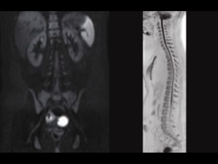
Whole Body Diffusion
Diffusion weighted imaging (DWI) is a powerful clinical tool for imaging patients. Thanks to advances in both hardware and software, Canon Medical’s MR systems offer excellent diffusion imaging quality. In combination with Olea Sphere, intravoxel incoherent motion (IVIM) for quantifying parameters that reflect tissue microcapillary perfusion and tissue diffusivity is now available in routine clinical practice.

MR Image Interpretation and Analysis
Olea Sphere allows the estimation of advanced qualitative and quantitative perfusion and diffusion parameters, providing instant access to multiparametric analysis as well as rendering and reporting tools to assist in diagnosis and communications.
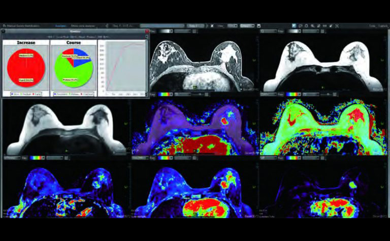
Breast MR
Breast MR is a highly sensitive method for imaging breast tissue.

Prostate Imaging
Canon Medical’s PURERF technology provides clinicians with the high resolution anatomical images required for MR imaging of the prostate. Multi b-value DWI, IVIM, and perfusion analysis can all be performed to assist with visualization, and all of them can be enhanced with the Olea Sphere Body package to provide a complete analysis package.
Time-SLIP / mASTAR
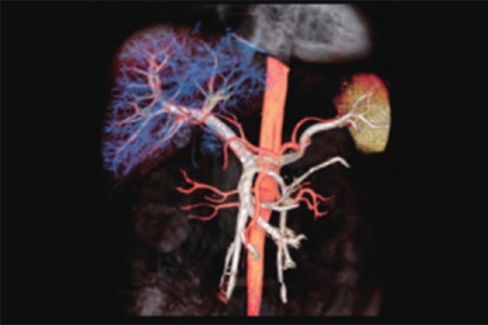
Time-SLIP technique may be helpful for preoperative imaging of the donor candidates for living-donor related liver transplantation. It may be helpful in imaging the following vascular variants: hepatic arterial variation, portal venous variation and biliary variants without contrast enhancement of MR. Time-SLIP may also be useful for imaging of Hepatic artery complications including Collateral Vessels before Transcatheter Arterial Chemoembolization and Radiofrequency ablation which are focal interventional procedures used to treat Hepatocellular Carcinoma.

Dynamic MR and Fat Fraction analysis by Vitrea
Dynamic MR and Fat Fraction analysis by Vitrea is useful in not only quantifying the perfusion metrics of hepatocellular carcinoma (HCC) and liver parenchyma, but also assessing perfusion changes after HCC chemoembolization. Angiogenesis is largely involved in the metastatic growth and progression of HCC, which is treated with transarterial chemoembolization (TACE) and systemic molecular targeted therapies.
The same kind of index can be produced using Dynamic contrast enhanced MR per below, requiring IV injection of contrast agents, and allows for the quantification of tissue and a tumor’s vascular characteristics. The Fat Fraction is helpful for diagnosis
with the DIXON MR Image.
Customer quote

“ My aim is to have the best image quality possible. New applications are explored with the latest developed sequences. With the Vantage Galan 3T, Canon Medical has a real competitive product offer in this market.”
Dr. Xavier Alomar
Head of Radiology
Clinica Creu Blanca, Barcelona, Spain