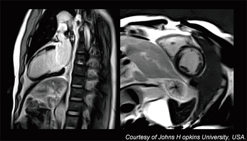
Achieve clinical freedom with Galan 3T
Vantage Galan’s fully digitized technology purifies the input and output signals which delivers sharper images. With unique Saturn Technology achieve enhanced signal to noise ratio (SNR) supporting diffusion weighted and high-resolution imaging.
Saturn Technology
Our intelligent Saturn Technology provides more consistent image quality through increased gradient stability and precise center frequency control.
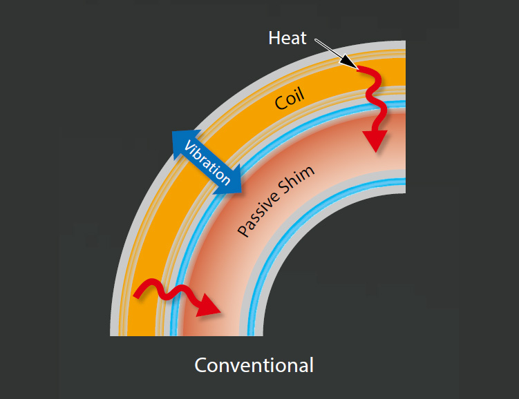
Increased gradient stability
With less vibration comes more stability resulting in crisper images. Saturn Technology delivers this through hardening the gradient coil with high-pressure molding. The result is less signal blur and thus better image resolution

precise center frequency control
In Vantage Galan 3T, increased image sharpness is achieved through improved thermal stability and thus a more stable center frequency. Triple cooling layers suppress temperature increases under high load leading to more stable image quality over long scan sessions.
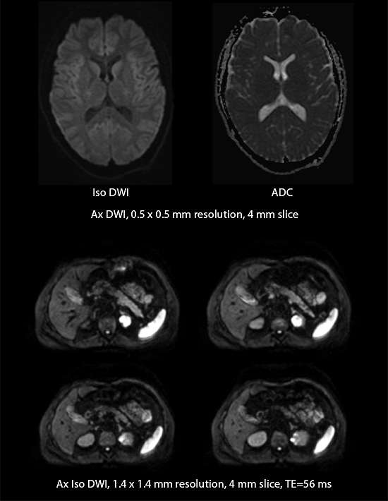
Diffusion Weighted Imaging With PUREGradient
With 45mT/m, 200T/m/s Saturn X Gradient performance, up to 30% increase in SNR in Diffusion Weighted Imaging in the brain and up to 51% in the liver can be achieved, resulting in enhanced diagnostic capability in diffusion imaging.
Pure digital signal for sharper images
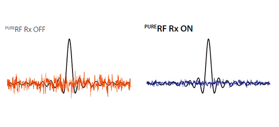
Our unique PURERF technology increases the SNR of Vantage Galan 3T by up to 20 percent for all clinical applications. The system’s digital RF transmit and receive efficiency enhances clinical confidence in imaging performance while shortening scan times.

ADVANCED POST PROCESSING ENHANCES DIAGNOSIS WHILE HELPING TO EXPAND PATIENT SERVICES
Advanced shielding design
Vantage Galan 3T’s unique PURERF Tx technology allows you to acquire sharper images with improved SNR. Its novel shielding design is aimed at maximizing the efficiency of RF transmission.
Advanced post processing enhances diagnosis while helping to expand patient services
Access advanced applications with Olea/Vitrea post processing tools
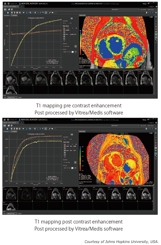
Full T1 map in a single breath hold with MOLLI
Expand your cardiac toolset with T1 mapping, allowing you to acquire a more quantitative characterization of myocardial tissue. T1 mapping utilizes a MOLLI sequence, enabling the acquisition of a full T1 map within a single breath hold.
Fewer breath holds with PSIR
Phase Sensitive Inversion Recovery (PSIR) in the heart provides improved contrast in late-enhanced imaging by using a more robust nulling of healthy myocardial signal without the need for an inversion time (TI) calibration scan. By eliminating the need for calibration, cardiac examinations can be completed with fewer breath holds and greater patient comfort.
[/inset]
