Automated bone subtraction and/or iodine mapping
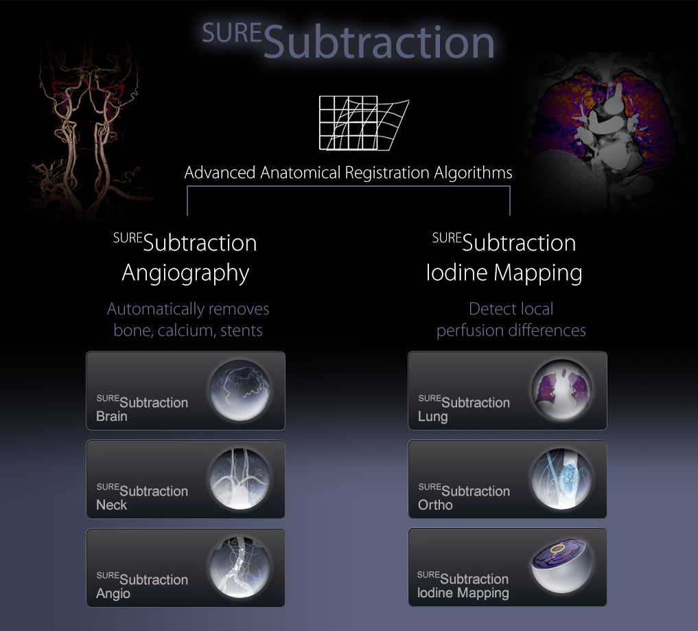
Canon Medical Systems’ SURESubtraction suite capitalizes on anatomically specific deformable registration algorithms to ensure accurate and robust results.
Angiography
- Automatically remove bone, calcium, stents
- Whole body CTA: Carotids, Aorta, Run-off, etc
- Automatic bowel removal
- Zero Click workflow
Iodine Mapping
- Visualize local perfusion differences
- Display local contrast enhancement
- Differential Enhancement Maps
- Virtual Contrast Boost
- Zero Click workflow


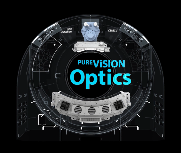
PUREViSION optics
GENESIS Edition transforms routine imaging to new levels of image detail and low contrast resolution – balanced for each clinical question at the right dose. A completely redesigned X-ray system from photon generation to beam distribution and detection is the basis of PUREViSION Optics. This results in a better balance between image quality and dose. Adaptive scatter correction removes scatter through intelligent modeling that preserves more primary photons for reconstruction as compared to a hardware-based approach.
The right balance between image quality and dose for every patient, from the youngest to the largest
- X-ray Generation – Small focal spot imaging for a wider range of exams
- X-ray Distribution – Adaptive Beam shaping optics ensure homogenous photon spread to maximize resolution for each clinical task while minimizing dose for patients of all shapes and sizes.
- X-ray Transmission – GENESIS utilizes raw data based scatter modeling and correction technology to ensure uniform image quality. A smart approach compared to scatter grids which sacrifice primary photons before they are ever detected.
- X-ray Detection – ThePUREVision Detector produces 40% more light to the photodiode. A breakthrough in scinilator production from a single praseodymium activated ceramic ingot.


Eliminating the workflow challenges of MBIR
Integrated, easy to use, and fast
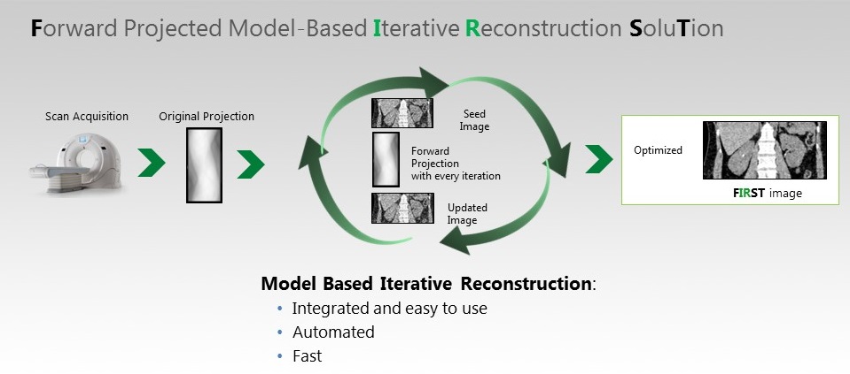
GENESIS Edition provides sharper image detail and lower patient dose with the world’s first fully integrated MBIR (Model-Based Iterative Reconstruction) solution.

FIRST utilizes forward projection iterations to deliver high-quality images with up to 82% dose reduction. A full volumetric reconstruction for routine clinical use can be obtained in just 3 minutes. Following Canon’s longstanding philosophy of minimizing dose while maintaining efficient clinical workflow, FIRST integrates seamlessly into your daily clinical practice.
Dynamic Volume CT – Simply efficient
More than a decade of clinical partnerships with leading institutions sets Canon apart as the industry leader in dynamic volume CT. Together, we have developed new procedures for better patient care, automated workflows, and refined reconstruction technology to make the remarkable routine.

PERFUSION
Transform your diagnostic capabilities from morphological to physiological diagnosis. Scans are performed effortlessly with low-dose parameters.
- Diagnose lesions that cannot be seen with traditional static imaging
- Add certainty to the classification of tumors with blood flow quantification
- Monitor tumor progression and follow-up response to treatment
- Reduce the need for additional imaging tests or biopsy
BODY PERFUSION
Body perfusion software analyzes the blood perfusion of an organ from dynamic scan images. The perfusion application can provide additional information for the diagnosis, treatment and follow-up of lesions.
- Whole-organ temporal uniformity
- Fully automated non-rigid volume position-matching software
- Low-dose perfusion scan protocol
- Interactive control of functional color overlay on morphological images

BRAIN PERFUSION
The Neuro ONE protocol allows acquisition of multiple low-dose volume scans of the entire brain during contrast infusion to provide whole-brain perfusion and whole-brain dynamic vascular analysis in one examination.

MYOCARDIAL PERFUSION
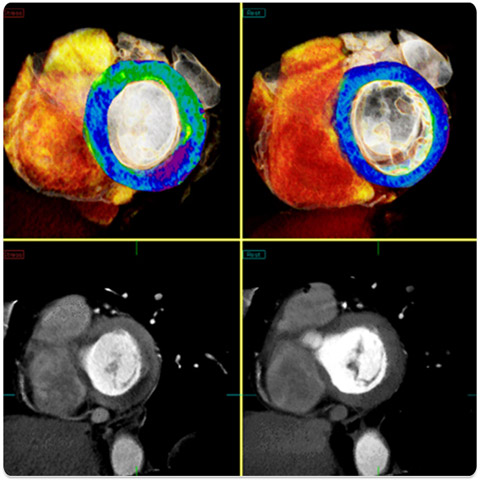
The results of the CorE320 multicenter trial have shown that coronary CTA has the potential to become a diagnostic tool equivalent to conventional coronary angiography. Following this trial, Myocardial Perfusion has been developed as an advanced application for quantitative perfusion analysis.
- Whole-heart acquisition in a single temporally uniform volume
- Accurate diagnosis: Myocardial Perfusion combined with coronary CTA can accurately assess myocardial perfusion and identify the coronary arteries responsible for ischemia
- Ultra-low dose: All required perfusion data can be obtained by just one scan
The right application for a confident diagnosis Automated, reliable, and robust
GENESIS Edition offers a comprehensive suite of Adaptive Diagnostic solutions to make complex exams easier and to improve diagnostic precision and reproducibility.

Subtraction CTA
Superior visualization in CTA with true subtraction of bone and calcification.

Iodine Mapping
Clearly defined perfusion with color blood flow maps as a result of advanced registration and subtraction.

SURECardioTM
The robust solution for coronary imaging with ONE shot volume imaging and arrhythmia scanning.

TAVR
Easily combined gated and non-gated acquisition for fast and low-dose TAVR exams.

SEMARTM
Improved visualization of bone and soft tissue – Single energy raw data based metal artifact reduction.

DE Tissue Characterization
Tissue Characterization with easy-to-use Dual Energy scanning.
