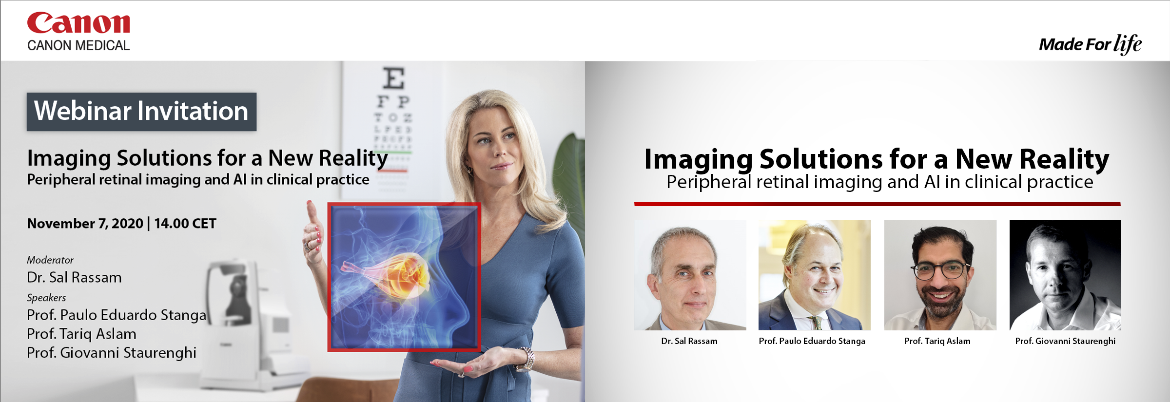
Webinar Eye Care
Imaging Solutions for a New Reality: Peripheral retinal imaging and AI in clinical practice
Background information: This unique event brings together renowned experts on retinal diseases and retinal imaging from all across Europe. They will address the unique product features that can change patient management. This webinar will increase your confidence while performing your daily routine with wide field OCT and AI functionality.
Learn from Prof. Paulo Stanga, Prof. Tariq Aslam and Prof. Giovanni Staurenghi how they use these devices in their daily practice to interpret the images and enhance their patient management.
Get practical tips from the examples demonstrated, to increase your diagnostic confidence.
Use this opportunity and participate in the live Q&A session lead by Dr. Sal Rassam to get the answers you need.
This webinar occurs now in the past. The recording is available. Click here to watch it now.
Watch again

Moderator: Dr. Sal Rassam, Worthing, UK
Dr. Sal Rassam is a consultant Ophthalmic Surgeon & Clinical Lead at Panacea Medical Centre Undertook a fellowship in disease of the Retina and Vitreoretinal Surgery at the Royal Victorian Eye and Ear Hospital in Melbourne, Australia and spent over 2 years conducting research on Ocular Circulation at the Hammersmith Hospital, London. . He has special interest in ocular circulation & ocular imaging.

Speaker: Prof. Paulo Eduardo Stanga, Consultant Ophthalmologist & Vitreoretinal Surgeon, London, UK
Title: First experience using Wide-Field Swept Source OCT
Abstract:
1. The Mid and Peripheral Vitreoretinal Interface and Neuroretina: Identification of normal anatomy and lesions
2. Vitreous Opacities, the Posterior Vitreous cortex and Posterior Vitreous Detachment: Objective assessment, staging and quantification
1. Successfully imaging the Mid and Peripheral Vitreoretinal Interface and Neuroretina can be considered a landmark achievement as it can potentially change clinical management and reduce the number of patients suffering from retinal detachment and needing surgery, by allowing the early recognition of lesions that need treatment or increase the risk of retinal detachment. Hence the significant importance of imaging the Mid and Peripheral Vitreoretinal Interface and Neuroretina and Posterior Vitreous Detachment.
2. Though generally accepted or dismissed by clinicians as non-clinically significant, Vitreous Floaters or Opacities are ubiquitous throughout human experience and generally considered innocuous. However, they can be debilitating for many people while impairing their quality-of-life due to degradation in contrast sensitivity function. Increasing use of backlit screens is emphasizing the magnitude and severity of this problem. With predicted 5 billion myopes worldwide by 2050, one can foresee an epidemic, since Myopic Vitreopathy is the second leading cause for clinically significant vitreous floaters: “Vision Degrading Myodesopsia”. Hence the significant importance of imaging Vitreous Opacities, the Posterior Vitreous cortex and Posterior Vitreous Detachment to objectively establish which patients may benefit from treatment and what the best choice of treatment for each patient may be; either YAG Laser Vitreolysis or Pars Plana Vitrectomy Surgery.
A selection of cases, treatment options and results will be presented and discussed.

Speaker: Prof. Tariq Aslam, Professor of Ophthalmology and Interface Technologies, University of Manchester, Manchester, UK
Consultant Ophthalmologist, Manchester Royal Eye Hospital, Manchester, UK and Director, IOVS MSc Course, Manchester, UK
Title: Applications of intelligent denoise in OCT-A
Abstract: In this presentation, I will be discussing the potential applications of the Canon OCT-A system from personal experience of its use in Manchester Royal Eye Hospital. I will cover clinical applications and use as well as some early data and potential applications for research use.
In terms of clinical applications, we have been utilising the Canon system for around a year at Manchester Royal Eye Hospital retinal clinics. We have in particular been exploring the ability of the Canon system to provide a wide-field OCT-A view that has been optimised using artificial intelligence technology. I will demonstrate the quality and review the validity of these artificial intelligence enhanced scans and display clinical scenarios where they have provided important information for patient management in medical retinal and uveitis patients.
In terms of research, the wide field enhanced scans provide new opportunities for detailed analysis of vascular structures. I will discuss current and projected projects exploring the utility of these scans and how such research applications may impact on patient care in the future.
Speaker: Prof. Giovanni Staurenghi, Chairman University Eye Clinic, Milan, Italy
Title: Wide Field OCT and OCTA
Abstract: Today the wide field imaging it is a indefeasible tool in a retina clinic. In particular the most recent imaging modalities which expanded their ability to a wider field are the OCT and the OCT-A.
First generation time-domain OCT devices were able to scan only a restricted portion of the posterior pole, with limited tissue penetration and relatively low resolution. Subsequent spectral-domain OCT technology allowed for a more detailed visualization of the posterior pole anatomy and enhanced view of deeper tissues. More recently, swept-source OCT has further enhanced penetration of the OCT signal, and with higher scanning speeds, has facilitated the acquisition of larger fields of view.
The steep curvature of highly myopic eyes can impact the visualization of the posterior segment on OCT. The tilted segments of the scan may impact the visibility of certain retinal structures. The size of the imaging window may also impact how much of the retina can be visualized in a normal orientation without conjugate image artifact impacting the more peripheral aspects of the scan.
The advantage of the SS-OCT is to have a bigger size of the imaging window (for the Canon Xephilio OCT-S1 5 mm).
Fluorescein angiography is an invasive test and it is useful in many conditions. However to identify the ischemic areas in diabetic retinopathy and retinal vascular occlusions OCT-A could much easily detect them due to the high contrast. Until now the limitation of the field of view did not allow the routinely use of this technology for the examination of the retina out of 20°. The introduction of the SS-OCTs and the possibility to create a montage allow a field of view of around 105-110°.