Visualize the microvasculature of the retina with OCT angiography
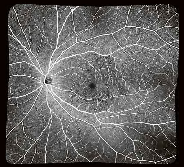
OCT angiography is a sophisticated technology that detects the movement of red blood cells in the retinal vasculature and allows you to visualize tiny vessels in detail.
Non-invasive examination, results within seconds
OCT Angio does not require fluorescein injection or pupil dilation, and the examination takes only seconds. SLO-based real-time tracking minimizes artefacts. Sophisticated image post-processing with 3D projection artefact removal enables excellent image quality.

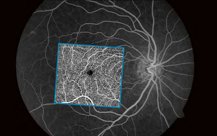
Angio Expert with freely selectable layers
With OCT angiography even the smallest blood vessels can be observed in 2D and 3D. With Canon’s OCT Angio software, you can freely select layers to create the preferred image. Layers can be defined based on automatic segmentation or as a custom offset.
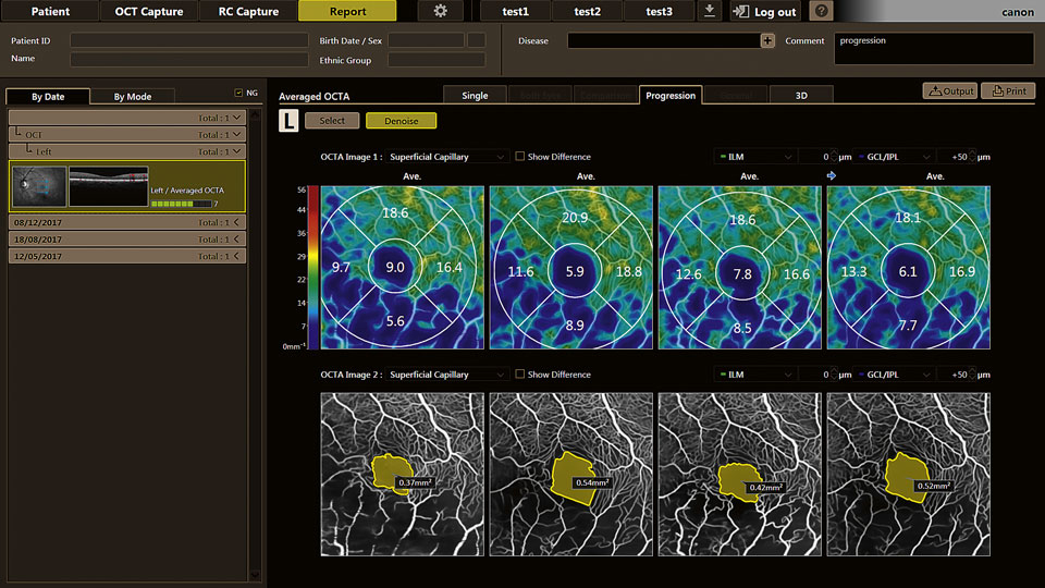
Analysis and reporting tools
Canon Medical’s Angio Expert software provides a complete set of manual and automated analysis tools, including distance, area, area density and skeleton density. The associated progression report displays up to four exams simultaneously next to each other.
Unlock the full potential with Angio Expert HD
Take advantage of the full potential of Xephilio OCT-A1 with the optional HD software. Angio Expert HD not only offers a wide range of advanced image quality tools, but also adds advanced OCTA analysis to your portfolio.
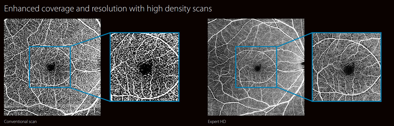
Angio Expert HD gives you a higher pixel density and an extended field of view, without losing the image resolution even from wide angles. In this way, you can image vessels and capillaries over a large area with high precision. While a standard scan has a size of 232 x 232 pixels, the HD-enabled high-density scans offer extended formats of up to 696 x 696 pixels to provide excellent image quality.
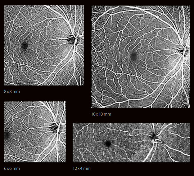
Always the right angle
With Angio Expert HD, you can choose the optimal scan density for any viewing angle you choose. The system provides various square and rectangular formats from 3 x 3 mm to 10 x 10 mm and 12 x 4 mm.
Panoramic imaging
With the optional Mosaic software, you can create ultrawide OCTA images up to 17.5 mm in length from 4 or 5 shots. Mosaic also lets you scan difficult-to-image patients in multiple sessions. It then uses faster but smaller scans, 6 x 6 mm which can be combined into scans of the required size.