High-definition imaging
Xephilio OCT-A1 offers excellent image quality due to our optical 3 micron axial / 1.6 micron digital resolution.
Vitreo-Retinal Traction
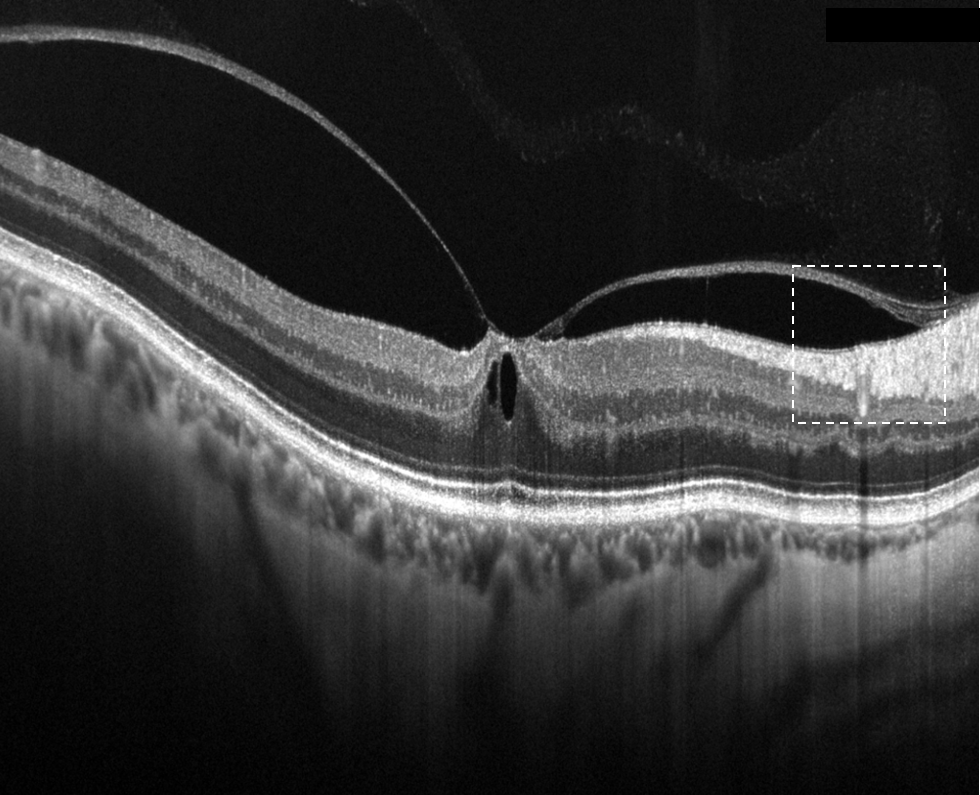
Courtesy of Adil EL MAFTOUHI, Centre Rabelais, Lyon, France
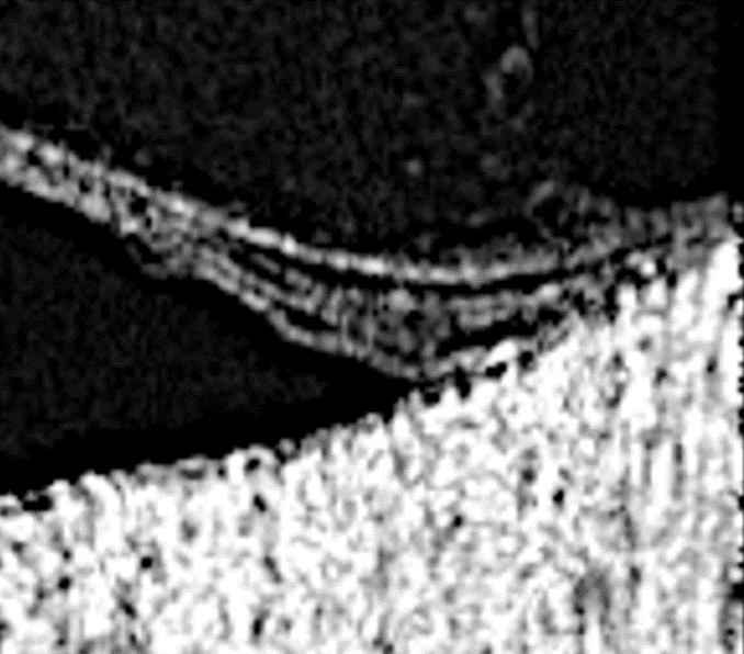
Partial magnification detail
Posterior Vitreal Detachment
Vitreo-Schisis in Early PVD associated with slight foveal traction
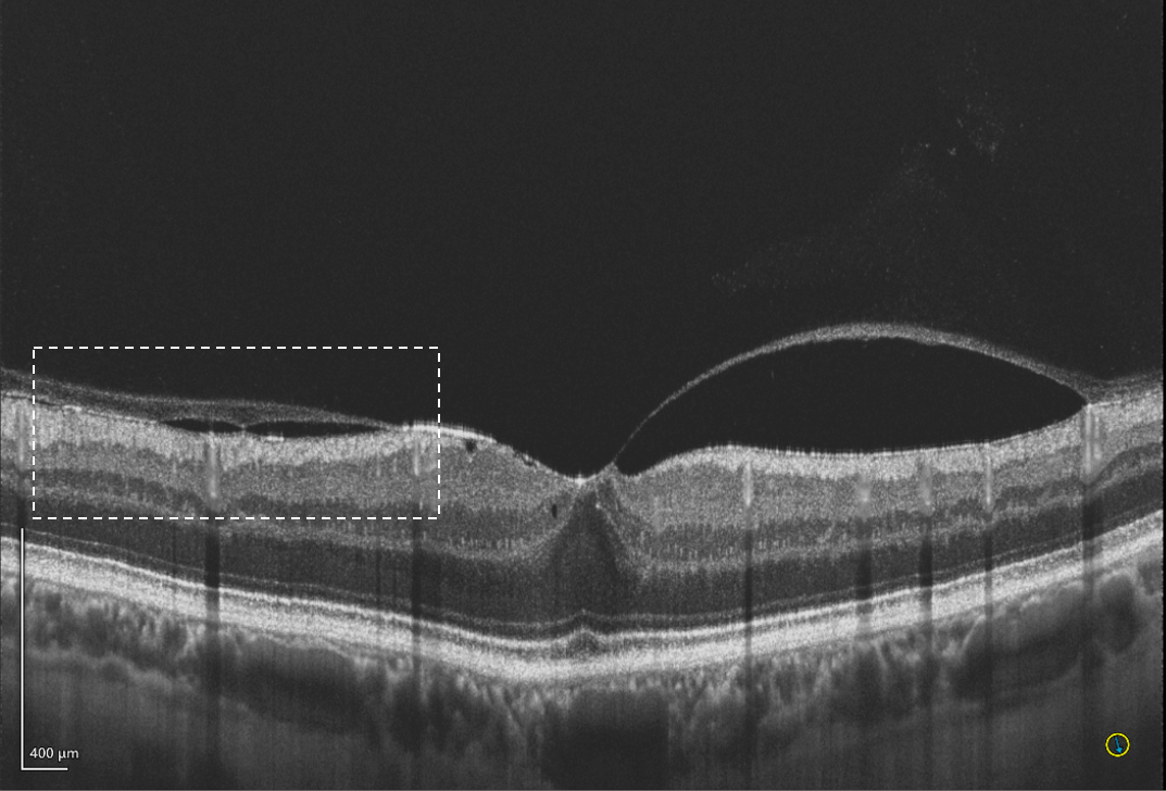
Courtesy of Adil EL MAFTOUHI, Centre Rabelais, Lyon, France
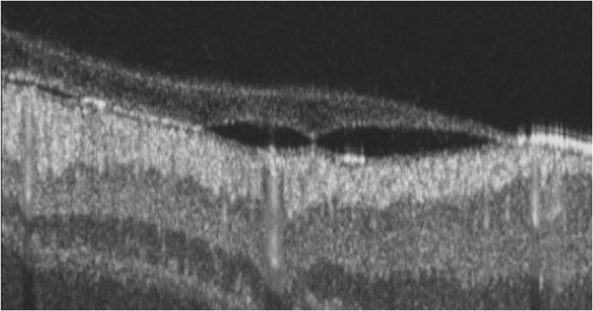
Partial magnification detail
Tear Film Analysis
Tear film analysis after Femto Lasik
Clear visualisation of the tear film above the epithelium
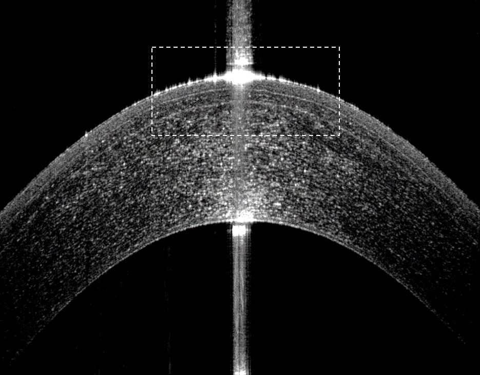
Courtesy of Adil EL MAFTOUHI, Centre Rabelais, Lyon, France

Partial magnification detail
Femto LASIK
Corneal Flap of Femto myopic lasik procedure with regular cutting.
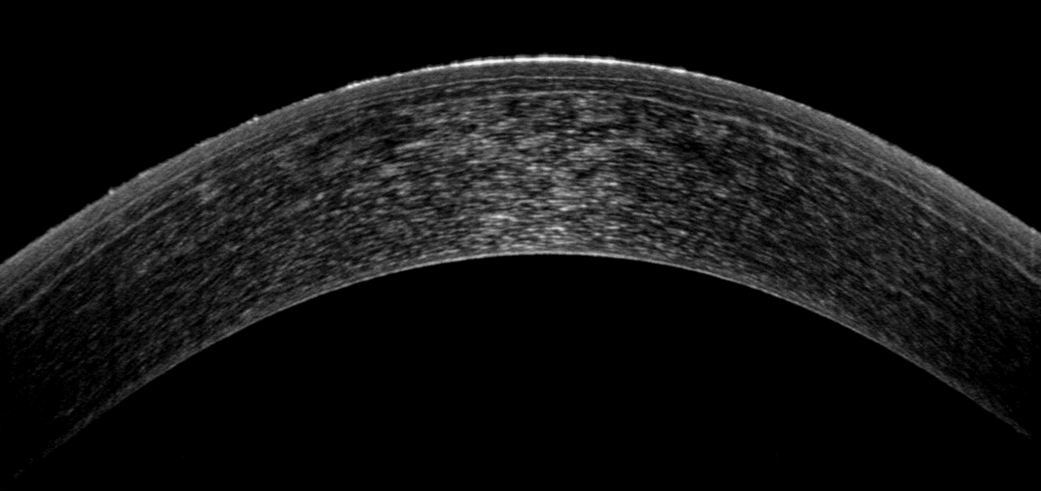
Courtesy of Adil EL MAFTOUHI, Centre Rabelais, Lyon, France
Hyperopic Lasik complication with Central Corneal Scar
History of Hyperopic lassie with a complication with Central and superficial opacities. Due to the optical resolution from Xephilio clearly shows the depth of the scar.
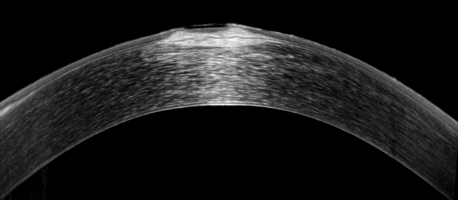
Courtesy of Adil EL MAFTOUHI, Centre Rabelais, Lyon, France
LASIK
History of lasik, 14 years ago, for myopic procedure (-4D) with microkeratome. Corneal flap is clearly visible in BSCAN. Micro ondulations of Basal membrane are highlighted due to micro folds of the corneal flap formed by the partial displacement of a lasik flap. This micro folds is responsible for the light streaks visualization.
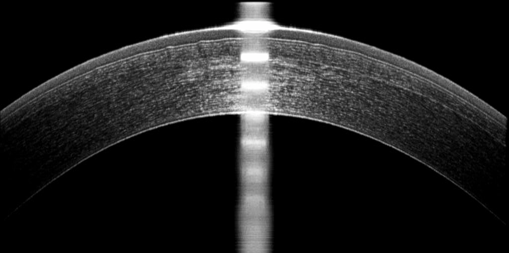
Courtesy of Adil EL MAFTOUHI, Centre Rabelais, Lyon, France
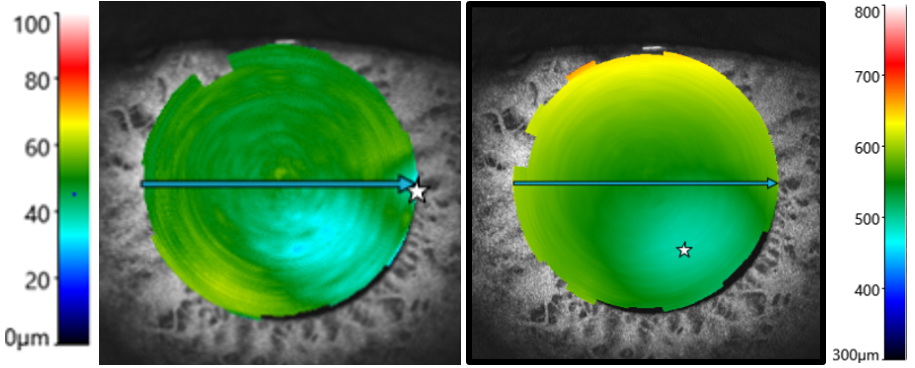
Epimapping and Keratoconus Screening
Bilateral keratoconus
Total Pachymetry shows a shift from the thinnest point with decentred thickness distribution in infero temporal part. Epithelium Mapping highlights clearly epithelium thinning in the cone area according to conventional corneal topography in both eyes. Epithelium Mapping is an additional tool for keratoconus screening.
Right eye

Corneal Thickness and Epithelium Thickness mapping

Left eye
Epithelium Thickness mapping and Corneal Thickness

Courtesy of Adil EL MAFTOUHI, Centre Rabelais, Lyon, France