Clinical cases
Xephilio OCT-S1 enables superior penetration of dense objects and provides outstanding tomographic images. Review here several clinical cases.
Retina
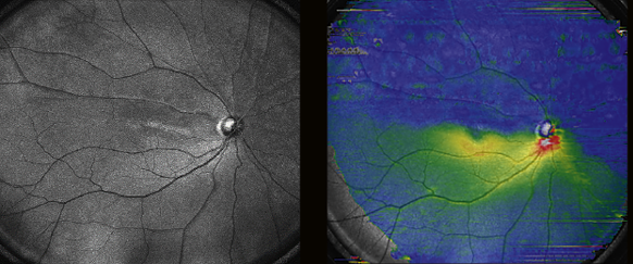
image C

image E
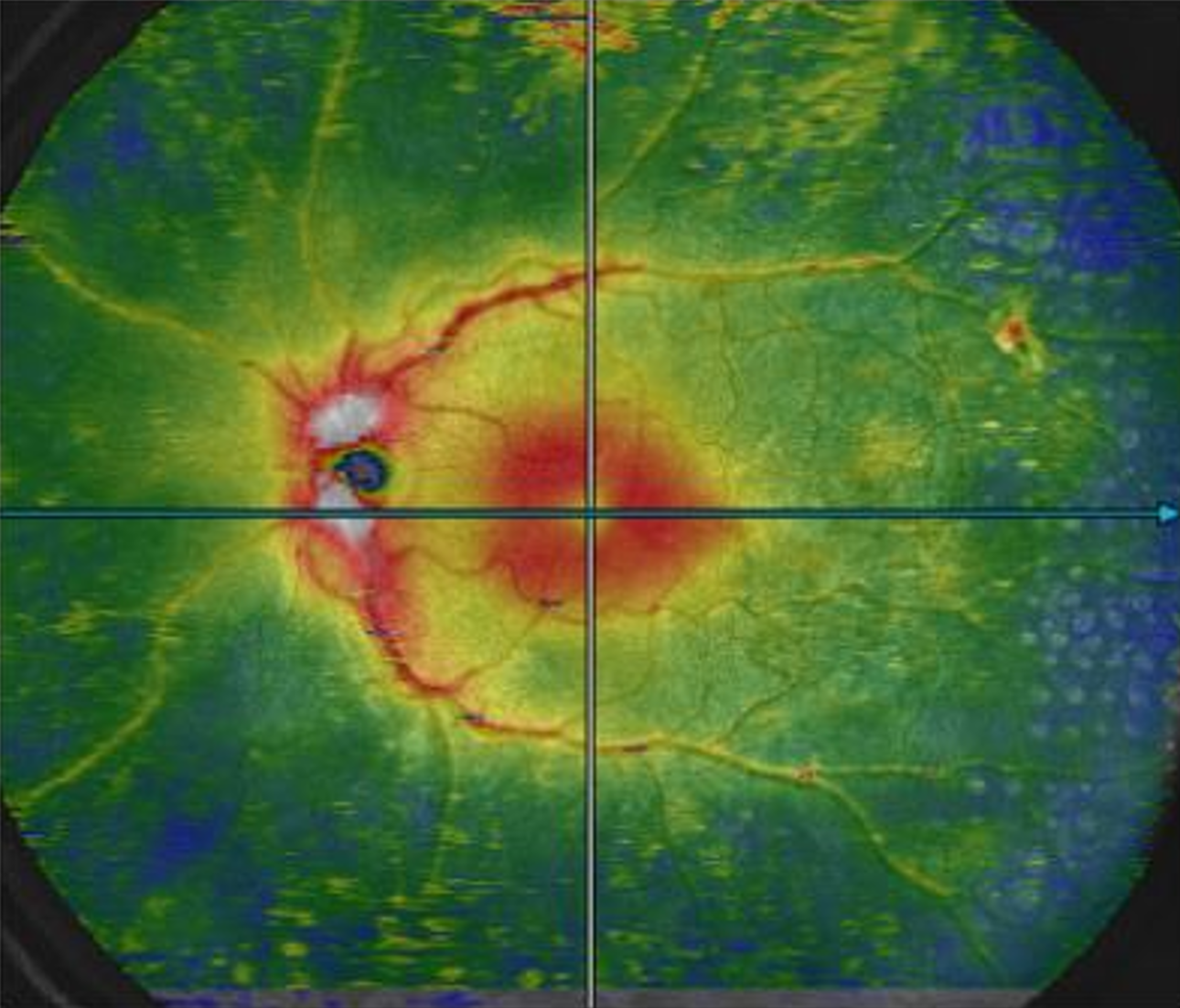
image A
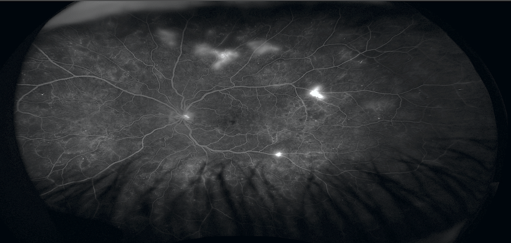
image B
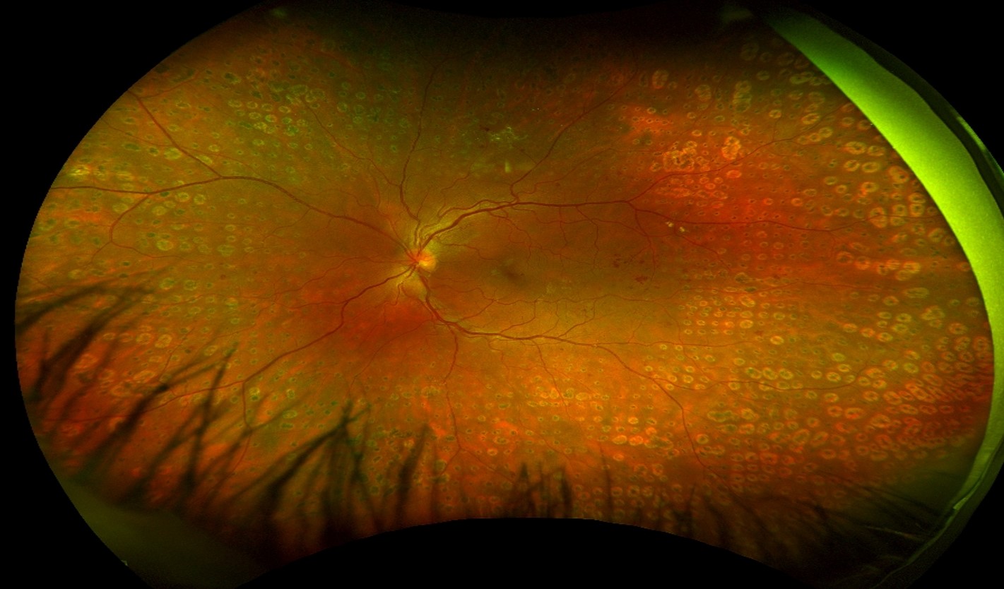
image D

C: Post Treatment Single-scan Wide-Field OCT Angiogram
Post Treatment Single-scan Wide-Field OCT Angiogram obtained on the same date as image B. Comparable in vertical axis extension to UWF-FFA on image A. Perfused and non-perfused retinal areas are clearly defined without the need of intravenous dye. Note that the residual fibrosed superior mid-peripheral NVE remains perfused and the residual minimally fibrosed temporal mid-peripheral NVE is perfused and not visible on image B.
A: Ultra Wide-Field Fundus Fluorescein Angiogram (UWF-FFA)
Ultra Wide-Field Fundus Fluorescein Angiogram (UWF-FFA) showing treatment-naïve active Proliferative Diabetic Retinopathy (PDR) with posterior pole and mid-peripheral neovascularisation elsewhere (NVE).
B: Ultra Wide-Field Multi-Wavelength Imaging
Ultra Wide-Field Multi-Wavelength Imaging after successful Targeted Retinal Photocoagulation (TRP) showing fibrosis and apparent complete resolution of superior and temporal mid-peripheral NVE.
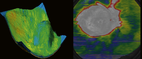
D: Post treatment macula-only OCT Retinal Thickness Map
Post treatment macula-only OCT Retinal Thickness Map obtained on same date as image B.
E: Wide-Field OCT Retinal Thickness Map
Wide-Field OCT Retinal Thickness Map obtained on the same date as images B, C and D. Note the mid-peripheral retinal thinning corresponding with areas of laser treatment and localised areas of thickening corresponding with the residual superior and temporal mid-peripheral NVE.
Courtesy of Prof. Paulo E. Stanga, Consultant Ophthalmologist & Vitreoretinal Surgeon, London Vision Clinic – Retina Lead, UK
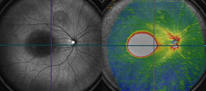
Branch retinal artery occlusion
Chronic central retinal vein occlusion

Rhegmatogenous retinal detachment

Central Serous Chorioretinopathy
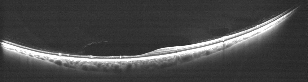
Anterior segment OCT*
Xephilio OCT-S1 allows you not only to visualize the microvasculature of the retina, but also of the conjunctiva. The anterior segment can be observed without the need for any additional lens attachments.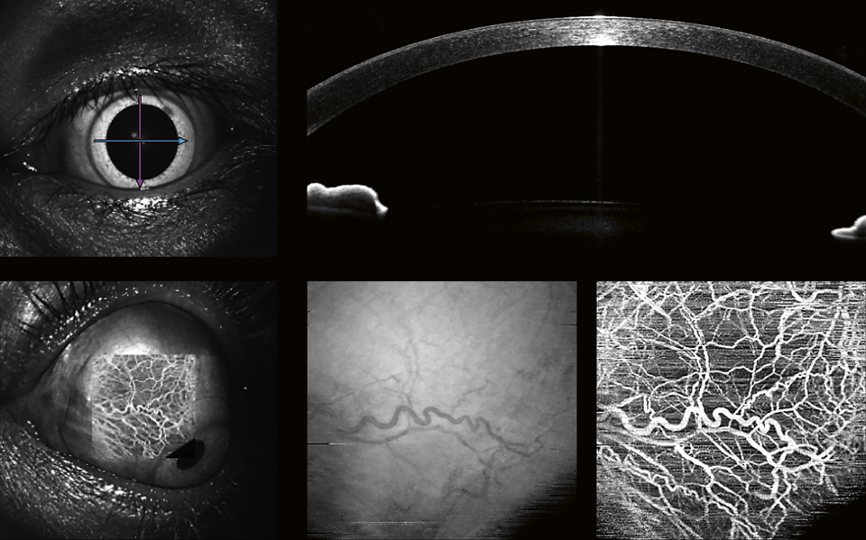 En-face OCT and OCTA
* Anterior segment OCT is currently intended for research purposes only and must not be used for patient diagnoses.
Intelligent denoise

Original image (w/o intelligent denoise)
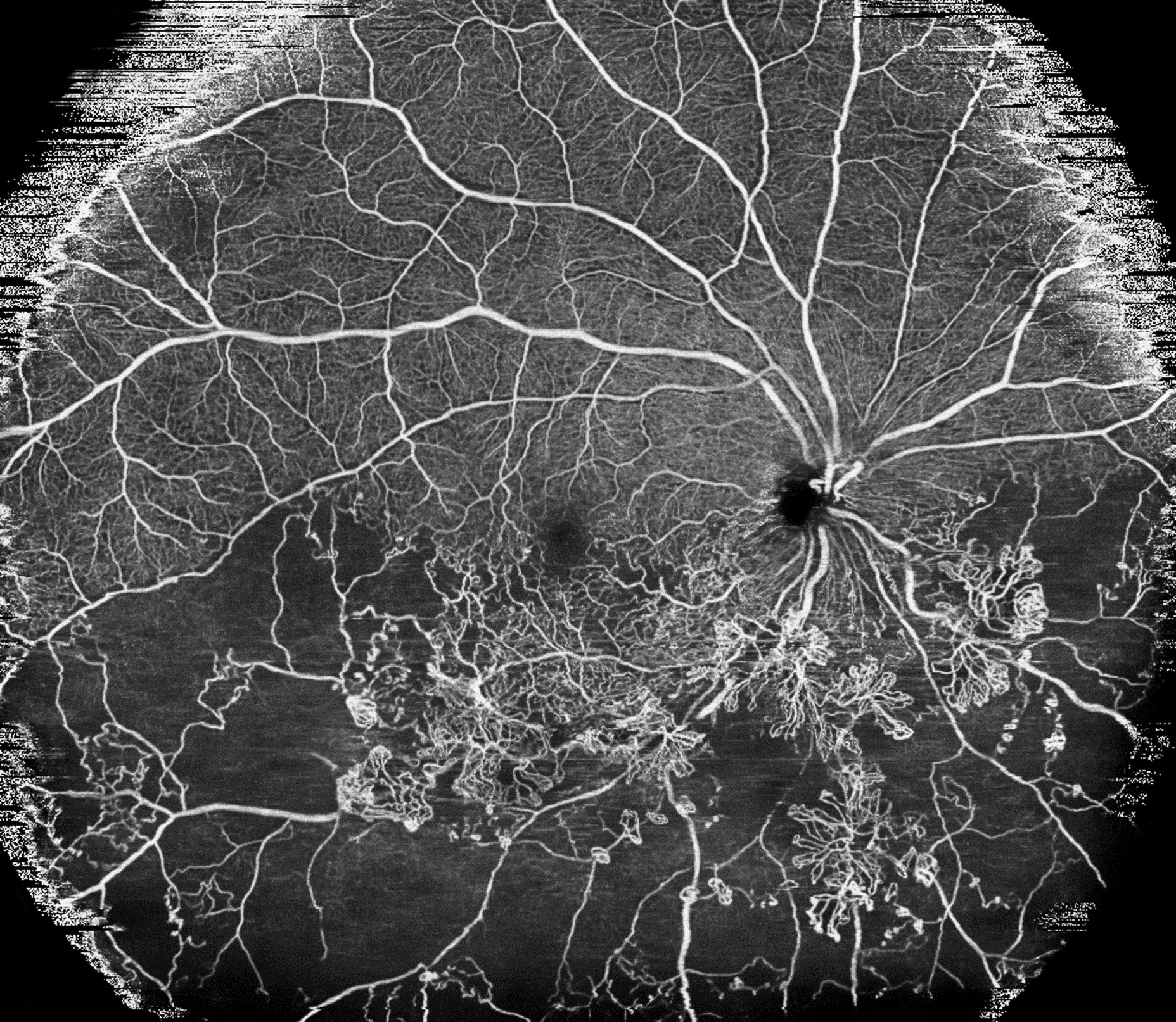
Image with intelligent denoise
Single scan wide field OCT Angiography
Post treatment single scan wide field OCT Angiogram. Perfused and non-perfused retinal areas are closely defined without the need of intravenous dye. Note that the residual fibrosed superior mid-peripheral NVE remains perfused and the residual minimally fibrosed temporal mid-peripheral NVE is perfused.
Courtesy of Prof. Paulo E. Stanga, London Vision Clinic, UK.
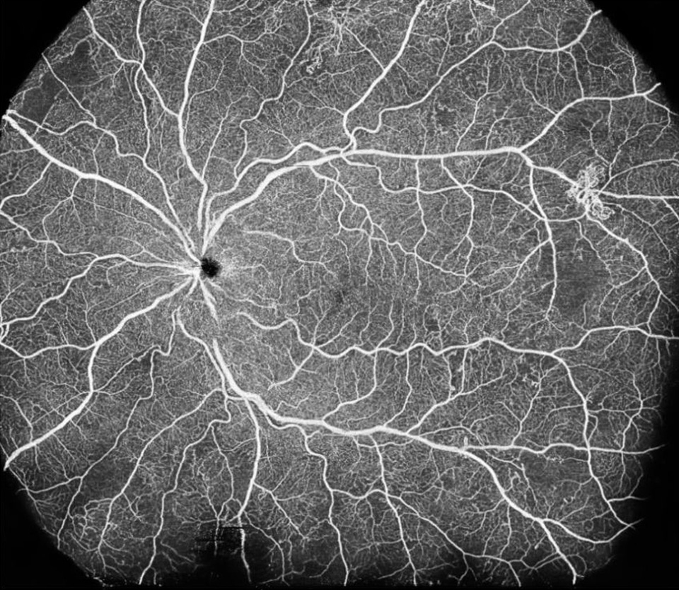
Wide Field En Face OCT
Xephilio OCT-S1 provides wide-field imaging of up to 23 ×20 mm width with just one scan. Mosaic imaging allows you to create a wide-field OCT image of approximately up to 31 x 27 mm with just 4 or 5 images.
Presentations during Congresses
Dr. Shin Kadomoto speaks about the impact of ultra-widefield swept source OCT and OCT Angiography … and its incredible field of view, during the OCT Congress in Rome, 2019.
https://youtu.be/6mSral4rsTM=rel=0
Dr. Lorenzo Casillo speaks about the everyday practice with the new Canon A1 SD-OCTA with OCT Angiography, during OCT Congress in Rome, 2019.
https://youtu.be/gXO4zUCzzzA=rel=0