Fast 3D Mode
”With Fast 3D, the scan time can be reduced while maintaining image quality. In addition, vessels are visualized as low-intensity areas, making it possible to more clearly discriminate between tumors and surrounding tissues. Since Fast 3D is relatively unaffected by magnetic field inhomogeneity, it is particularly useful for detecting lesions in the skull base or the posterior cranial fossa.
In this case, an irregular mass exhibiting contrast enhancement is observed in the right orbit. In the image acquired using FFE 3D, the lesion cannot be clearly visualized due to artifacts caused by magnetic field inhomogeneity. On the other hand, in the image acquired using Fast 3D, the orbital tumor can be clearly visualized. In addition, the scan time is shorter with Fast 3D as compared to FSE 3D.”
– Dr. Kohei Hamamoto, Jichi Medical University Saitama Medical Center, Japan

k-t SPEEDER
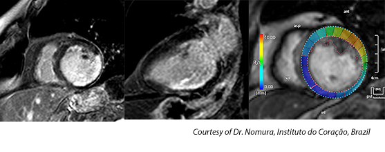
“Cardiomyopathy revealed severe systolic dysfunction on K-t speeder cine images. There was full-thickness late gadolinium enhancement of the entire inferior wall, inferolateral walls of the left ventricle”
– Dr. César Nomura, Instituto do Coração, Brazil
– Dr. César Nomura, Instituto do Coração, Brazil
MultiBand SPEEDER
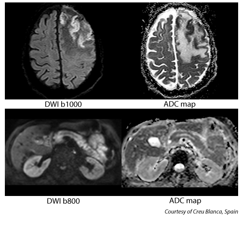
“MultiBand Diffusion Sequence has reduced our clinical scan times by half compared to the traditional Spin Echo Diffusion for both Brain and Abdominal Regions. Moreover, the new multiband sequence has helped reduce artifacts in areas of high susceptibility such as air and bone while overall improving ADC maps.”
– Dr. Xavier Alomar, Creu Blanca Medical Center, Spain
– Dr. Xavier Alomar, Creu Blanca Medical Center, Spain
Olea NOVA+™
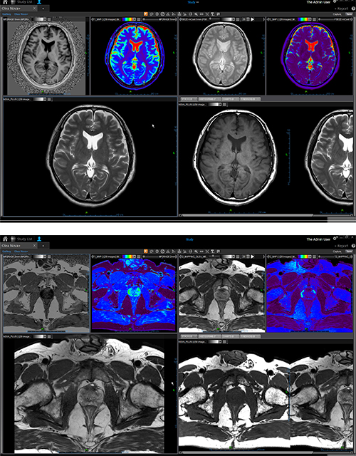
“Olea Nova+ allows for the Realtime tissue characterization of lesions in the prostate by only changing the calculated TE value on Olea Sphere. Olea Nova Plus of the prostate enhances the clinical confidence since the calculated TE values/images can be calculated retrospectively. This New Clinical feature from Canon Medical changes the way of evaluating cancers of the prostate while reducing the overall examination time.”
-Dr. Xavier Alomar, Creu Blanca Medical Center, Spain
-Dr. Xavier Alomar, Creu Blanca Medical Center, Spain
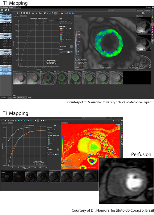
T1 Mapping
“In cardiac MRI, T1 mapping allows us to evaluate the myocardial tissue properties quantitatively. Even for diffuse lesion that are difficult to be evaluated with LGE, we can evaluate in absolute value with T1 mapping. Therefore, it contributes to the improvement of the diagnostic ability and give additional values to the image diagnosis.”
-Akiyuki Kotoku M.D.,Ph.D, Department of Advanced Biomedical Imaging, St. Marianna University School of Medicine, Japan
-Akiyuki Kotoku M.D.,Ph.D, Department of Advanced Biomedical Imaging, St. Marianna University School of Medicine, Japan
“T1 Mapping allows us to identify the pathologic area without the injection of contrast. In general, a prolonged native myocardial T1 signal is encountered in various disease states that result in edema or fibrosis, and in amyloid deposition”
-Dr. César Nomura, Instituto do Coração, Brazil
Wall Motion Tracking
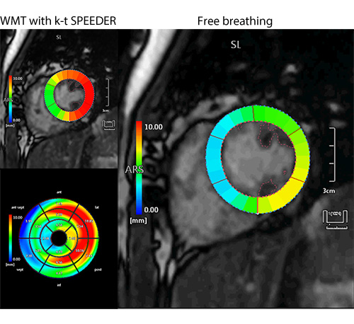
”With Wall Motion Tracking, local wall motion abnormalities associated with ischemia or cardiomyopathy can be evaluated using automatically calculated myocardial strain values. The presence of cardiac motion dyssynchrony, which is an indication for cardiac resynchronization therapy, can also be detected by analyzing multiple myocardial strain curves. In addition, global strain, which is a useful index for prognostic assessment and treatment planning in patients with cardiac failure, can be calculated automatically using both short-axis and long-axis images.
This software allows advanced cardiac function analysis to be performed using cine images acquired in routine examinations, eliminating the need to perform additional scans using special sequences. Retrospective analysis is also supported, making the software even more useful. In addition, the software permits cardiac contours, including the contours of the left ventricular papillary muscles, to be automatically traced with high accuracy, providing results with excellent reproducibility. Finally, analysis is performed very quickly, which helps to improve workflow.”
– Dr. Michinobu Nagao, Department of Diagnostic Imaging and Nuclear Medicine, Tokyo Women’s Medical University, Japan
