Superior penetration of dense objects
Revolutionary swept source technology allows you to capture wide-field images of up to 23 mm in a single scan. Xephilio OCT-S1 enables superior penetration of dense objects and provides outstanding tomographic images.

WIDE FIELD SWEPT SOURCE OCT IN ONE SINGLE CAPTURE
With Xephilio OCT-S1 Canon introduces revolutionary swept source technology allowing you to capture wide-field images of up to 23 mm in a single scan. Xephilio OCT-S1 enables superior penetration of dense objects and provides outstanding tomographic images. Experience a new quality of OCTA images in a single scan with greatly reduced noise, increased detail and improved visibility within just seconds.
AI-powered performance
AI helps you save time and improve imaging
Canon’s Intelligent Denoise, the system’s Deep Learning AI technology, offers a new quality of OCTA images based on individual scans – without the need to acquire and merge multiple images. The revolutionary technology delivers images with greatly reduced image noise, increased detail and improved visibility within just seconds.
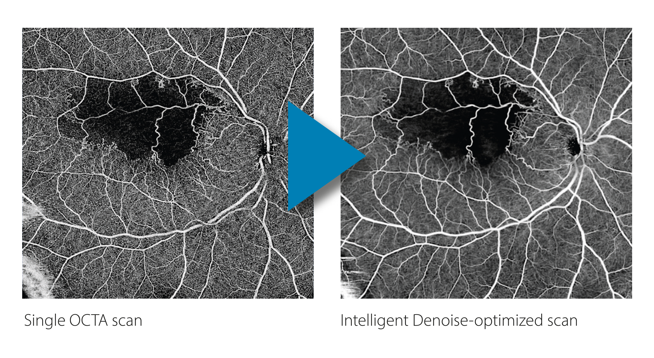
Courtesy of Centre Explore Vision, Paris, Dr. Maté Streho and Mrs. Néda Abraham.
Faster
100,000 A-scans per second combined with invisible 1,060 nm wavelength provide ultra-fast swept source technology maximizing data quantity of the patient’s eye while reducing acquisition time. Invisible scan lines ensure better patient collaboration and reduce the impact of patient eye movements.
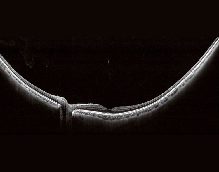
Deeper
Canon’s deep scanning swept source technology allows better penetration of cataracts, hemorrhages, blood vessels and sclera and at the same time optimizes capture of retinal and choroidal data – all in a single shot. With Xephilio OCT-S1 vitreous body and choroid appear in the same image with superior image quality providing more information for better patient care.
Wider
With a single capture the swept source Xephilio OCT-S1 shows a large wide-field OCT image as wide as 23 x 20 mm, which can be very beneficial for retina thickness observation of retinal detachment or retinitis pigmentosa. Mosaic imaging allows you to create an incredible 31 x 27 mm wide field OCT image.
 23 mm wide-field OCT B-scan image
 31 x 27 mm OCTA mosaic image
 Wide en-face OCT mosaic image