Visualize the microvasculature of the retina with OCT angiography
OCT angiography is a sophisticated technology that detects the movement of red blood cells in the retinal vasculature and allows you to visualize tiny vessels in detail.
Non-invasive examination, results within seconds
OCT Angio does not require fluorescein injection or pupil dilation, and the examination takes only seconds. SLO-based real-time tracking minimizes artefacts. Sophisticated image post-processing with 3D projection artefact removal enables excellent image quality.
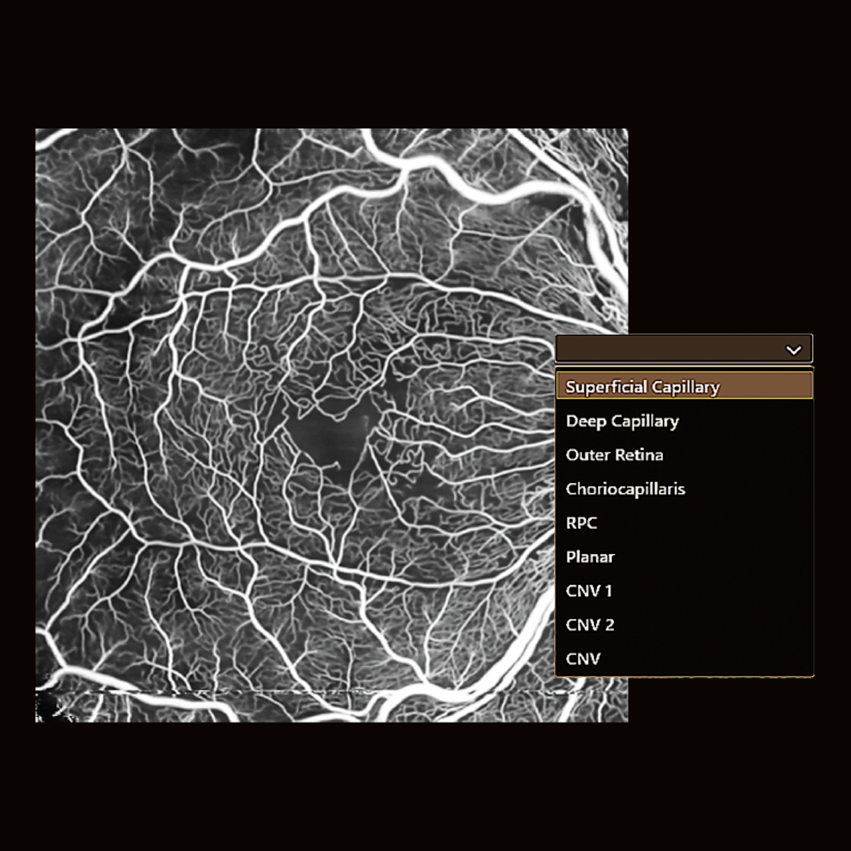
Angio Expert with freely selectable layers
With OCT angiography even the smallest blood vessels can be observed in 2D and 3D. With Canon’s OCT Angio software, you can freely select layers to create the preferred image. Layers can be defined based on automatic segmentation or as a custom offset.
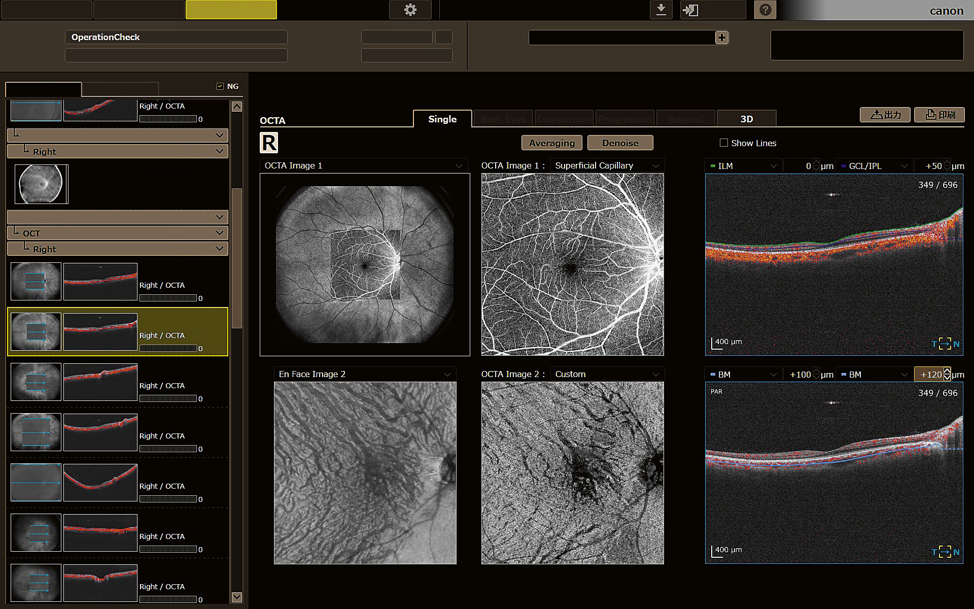
Analysis and reporting tools
Canon Medical’s Angio Expert software provides a comprehensive set of manual and automated analyses.
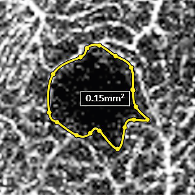
Automated area analysis and measurement
With a simple click on a non-perfused area or the foveal avascular zone, the target area is automatically detected, analyzed and displayed. If needed, users can change the automatically drawn borders or trace the area completely manually.
High density and single capture
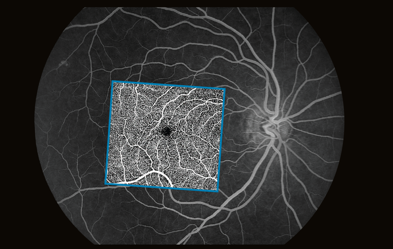
The Xephilio OCT-S1 offers an enormous diversity of scan areas and scan densities for OCT Angiography examinations. While scan areas range from small (3 x 3 ~ 8 x 8 mm) to super large (23 x 20 mm), a high scan density of up to 928 x 807 pixels allows for visualization of small vessels at the same time.

Always the right angle
With Angio Expert, you can choose the optimal scan density for any viewing angle. The system provides various square and rectangular formats from 3 x 3 mm to 23 x 20 mm.

Single capture wide-field imaging
Single scan wide-field imaging enables OCT Angiography of up to 23 mm width.This allows wide-spread non-perfused areas to be visualized which is useful in diagnosing diabetic retinopathy and retinal vein occlusion. At the same time, a single high-density
OCTA scan visualizes even small capillaries.

Panoramic imaging
The optional Mosaic software allows you to create ultra-wide OCTA images of approximately up to 31 x 27 mm.
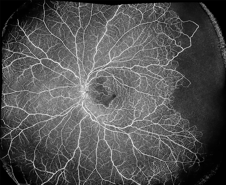
Central retinal vein occlusion. Courtesy of Prof Staurenghi, Sacco University, Italy