Outstanding imaging is your best friend
Canon’s recognized optical expertise enables the Xephilio OCT-S1 to offer superb image quality with minimal scatter. The swept source technology results in enhanced penetration further into the deeper tissue structures such as the choroid and even the sclera. Imaging depths of up to 5.3 mm allows for detailed visualization of the vitreous body and choroid in a single scan while the high scanning speed of 100,000 A-scans/s reduces examination time and offers very high resolution scans.

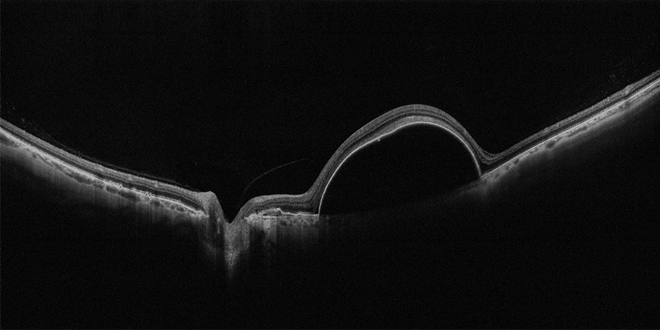
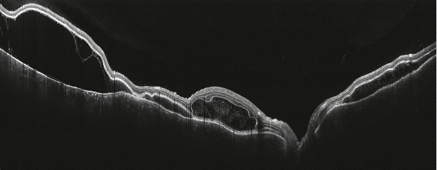
Serous PED. Courtesy Prof Staurenghi, Sacco University, Italy
Wider and deeper
With Xephilio OCT-S1 wide-field images of up to 23 mm width can be acquired in just one scan, equaling an 80° viewing angle. The 5.3 mm depth allows for visualization of the vitreous body and choroid in a single scan with superior image quality.
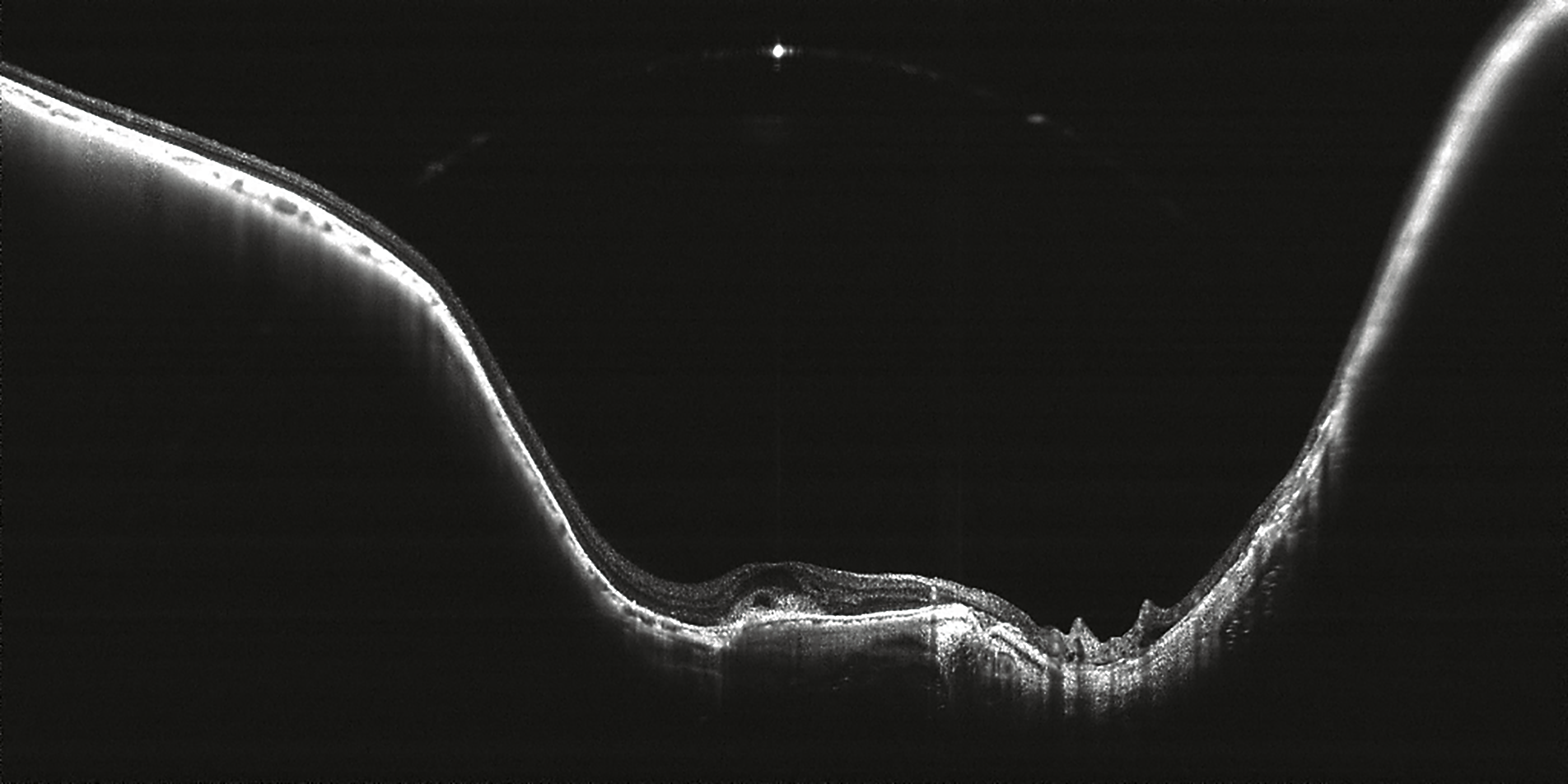
This 23 mm wide angle scan nicely depicts a chronic central retinal vein occlusion with edema.
Courtesy of Dr. Kadomoto, Kyoto University, Japan
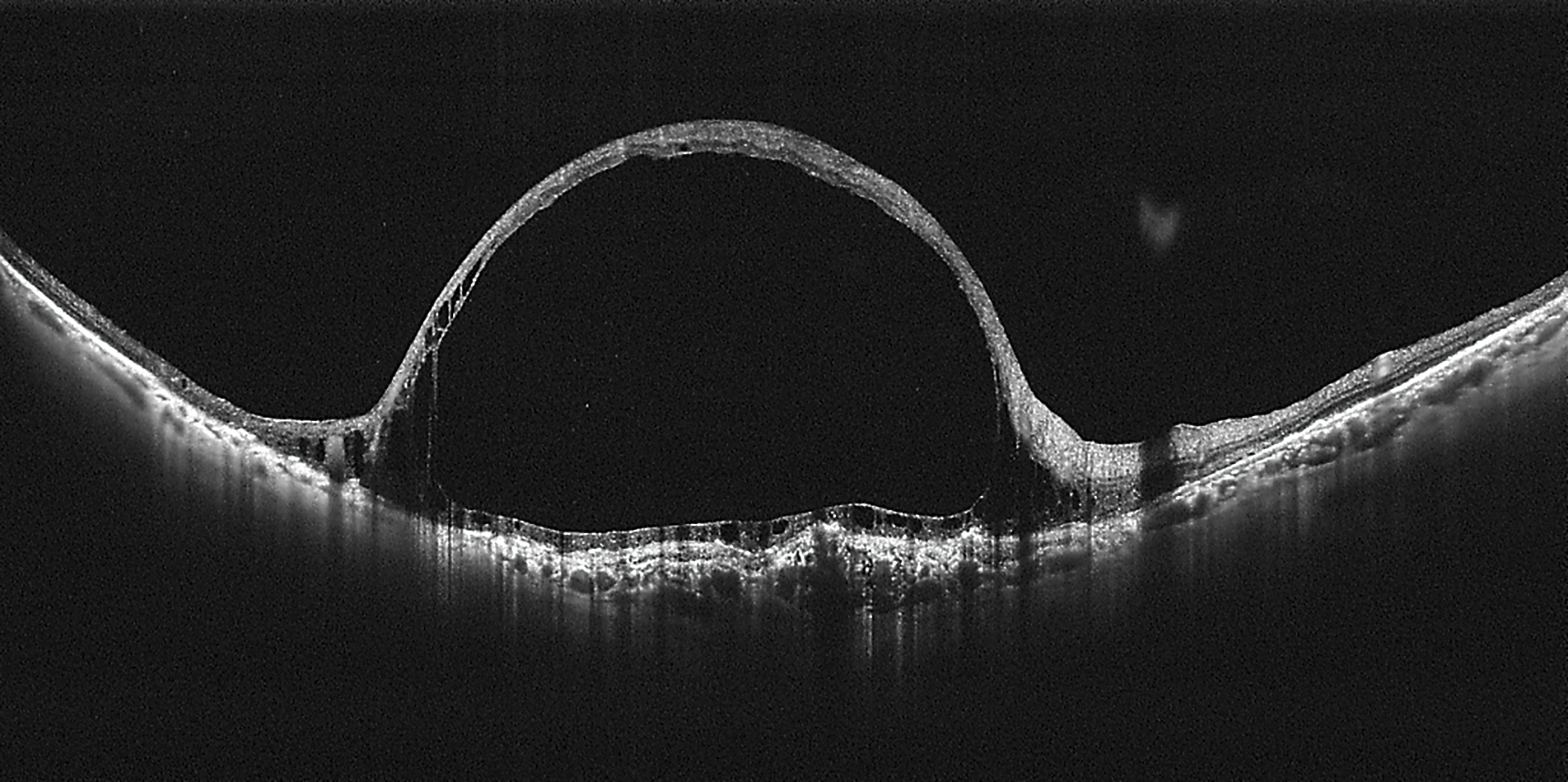
The curvature of the retina (especially posterior staphyloma) is well visualized in this Myopic Choroidal Neovascularization (mCNV) thanks to the 5.3 mm scan depth.
Courtesy of Dr. Kadomoto, Kyoto University, Japan
Single capture wide-field OCT
Xephilio OCT-S1 provides wide-field imaging of up to 23 ×20 mm width with just one scan. Mosaic imaging allows you to create a wide-field OCT image of approximately up to 31 x 27 mm with just 4 or 5 images.
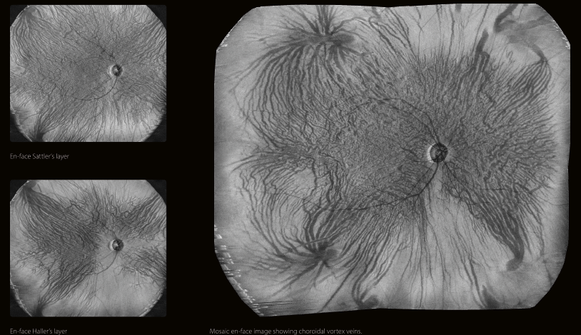
En-face Sattler’s layer, En-face Haller’s layer and Mosaic en-face image showing choroidal vortex veins.
Easy and quick operation
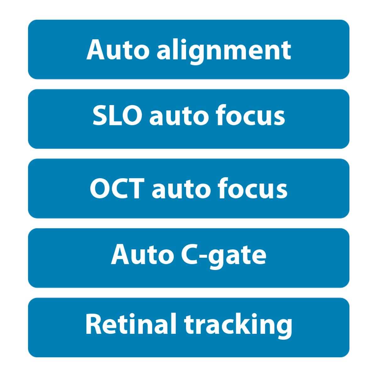
The Xephilio OCT-S1 utilizes a joystick for initial anterior alignment, but the operation is also aided by several automated functions. It has built-in SLO for real-time retinal tracking and accurate follow-ups.

The system’s joystick provides easy, quick operation combined with
pin-point precision.

The built-in optimization function automatically takes care of
alignment, focus and C-gate.
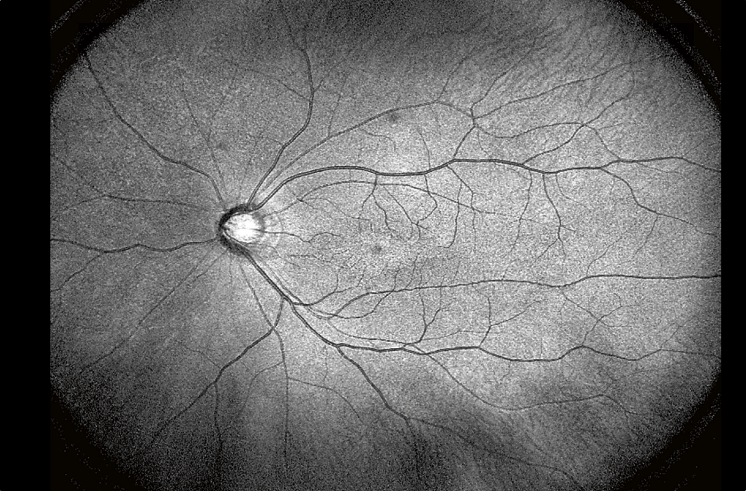
The wide-field SLO images acquired with Xephilio OCT-S1 allow for
superior observation.
Versatile reporting possibilities
Xephilio OCT-S1 provides you with a comprehensive range of reporting tools. Thanks to its extensive DICOM and EMR capability, results can be stored, shared and analyzed as needed in your daily practice.

3D reporting

OCT-A reporting
Outstanding imaging.
With Xephilio OCT-S1 Canon introduces revolutionary swept source technology allowing you to capture wide-field images of up to 23 mm in a single scan. Xephilio OCT-S1 enables superior penetration of dense objects and provides outstanding tomographic images.
Vogt–Koyanagi–Harada disease. Courtesy of Kyushu University Hospital, Japan